My Top 5 Coolest Studies of 2018!
Merry Christmas and Happy Holidays to everyone! I hope you all have a great new year :) I thought it would be fun to share my top 5 coolest studies of 2018 to round out the year! This is not a list of the ‘best’ studies of the year, as that is extremely hard to quantify (although all these are pretty stellar), so they are in no particular order. This is simply a list of papers that I thought tackled some interesting problems in a novel or unique way that made me think ‘damn that’s cool’. I hope you enjoy checking them out as much as I did! Click the paper titles for direct access to them.
#5 - Recovery of “lost” infant memories in mice
The phenomenon of “infantile amnesia”, where memories from early life are rapidly lost during development, has been known for quite some time. For example, almost no one has clear memories from when they were very little (e.g., 2 years old). Is this due to a problem in memory storage at this time…or are the memories stored properly, and we just can’t ‘retrieve’ them once we are adults? To investigate this question Guskjolen & colleagues used a transgenic approach to ‘optogenetically tag’ hippocampal neurons activated during the formation of an early fear memory in mice. Then, once the mice had grown up and ‘forgotten the fearful memory’, they reactivated those cells to see if they could ‘recover’ the lost memory. Indeed, they were able to do so suggesting that infantile amnesia is likely a result of retrieval failure rather than storage failure.
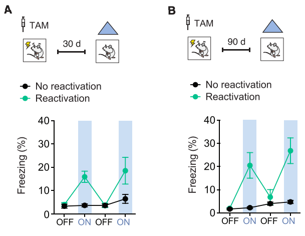
Reactivation of ‘lost’ memories in mice via optogenetic stimulation of neurons that were active during memory formation in early life. (A) Stimulation of cells activated in early life 30 days later caused mice to ‘freeze’, indicating they remembered the fearful memory; (B) this effect was long lasting, up to 90 days (longest they tested) (Credit: Guskjolen et al., 2018).
#4 - The neuronal gene arc encodes a repurposed retrotransposon gag protein that mediates intercellular rna transfer
Every once in a while there’s a paper that seems to turn biology on its head. This is an example of one of those papers, where the authors show that neurons can exchange transcriptional (RNA) information via secretion of ‘virus-like’ capsules composed of proteins thought only to be important in synaptic plasticity and memory formation. The proteins encoded by the gene arc seemed to form ‘virus-like’ structures that were able to travel between cells to exchange RNA information. This is because the gene that encodes these proteins shares an ancestry with those that made up ancient retroviruses! This type of inter-cellular communication has never been described (in mammals) and opens up a completely new regulatory and signaling pathway that may be important in neurodegenerative disease.

Intercellular transfer of messenger RNA via arc-encoded virus like proteins! (Credit: Pastuzyn et al., 2018)
#3 - Parallel circuits from the bed nuclei of stria terminalis to the lateral hypothalamus drive opposing emotional states
Ok I had to throw in this cool study out of my lab spearheaded by the great Will Giardino! Hypocretin/orexin neurons in the lateral hypothalamus modulate positive and negative aspects of arousal (e.g., promoting arousal in both rewarding, pleasurable, and stressful conditions). How a single neural population does this is unclear, but likely depends on differential inputs arriving from other brain areas. Giardino & colleagues demonstrated that different subsets of neurons in the bed nuclei of stria terminalis (BNST; extended amygdala) send projections to synapse onto hypocretin/orexin neurons, resulting in opposing responses depending on emotional state (positive or negative)!

Different populations of neurons in the BNST respond to positive (e.g., female mouse scent) or negative (e.g., predator odor) emotional stimuli . Time zero is when the stimulus was presented to the test mouse. On the Y axis you can see the fluorescent signal from CRF or CCK neurons in the BNST during stimulus presentation (credit: Giardino et al., 2018).
#2 - in toto imaging and reconstruction of post-implantation mouse development at the single cell level
Can we image every cell as a mouse develops to understand how a jumble of cells coordinates to make a complex organism like a mouse? Turns out we can! This one is just really damn cool. Check out the video below with a better explanation than I could ever give.
#1 - expanding the optogenetics toolkit with topological inversion of rhodopsins
What if we flipped the excitatory optogenetic protein channelrhodopsin upside down? Turns out it creates a pretty potent inhibitor of neural activity! I include this because it is such a simple idea, that turned out way better than I would have thought it could!…and the new opsin is called “FLInChR” which I thought was funny.

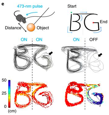
BOnus! - Medial preoptic circuit induces hunting-like actions to target objects and prey
How do animals engage appropriate behaviors necessary to survive, like stalking, hunting and chasing prey? Park & colleagues discovered that neurons in the medial preoptic area of the hypothalamus projecting to the ventral periaqueductal gray in the midbrain promote these behaviors in mice. Activation of this circuit (MPA—>vPAG) caused mice to chase, leap after, and hunt inanimate objects! Make sure to check out the figure and video link below to see this hunting behavior in action!

Activation of the MPA—>vPAG circuit promoted hunting-like behavior in mice. Here, the researchers drew (with a little ball on a stick) the letters “B” and “G”. When the laser was off, mice were scared of the object and stayed towards the edge of the arena, but when the laser was on, they hunted the object, so closely that they essentially drew the letters with their body chasing the ball!
There were many more studies that I wanted to include on this list…but I thought 5 was a good number to shoot for as 10 would have been too much! Maybe I’ll do another list for the most ‘impactful’ studies of 2018…but that will have to wait for another time (as it takes time to assess impact! All the best and happy holidays!! —JCB